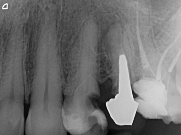
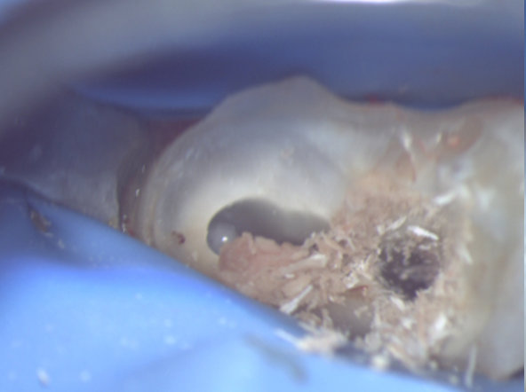
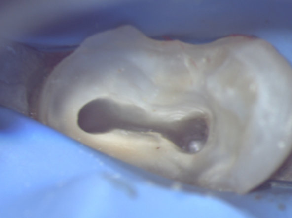
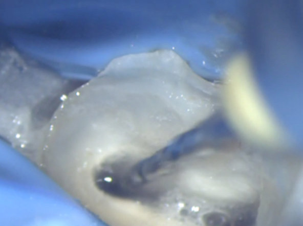
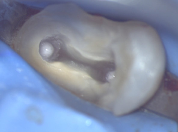
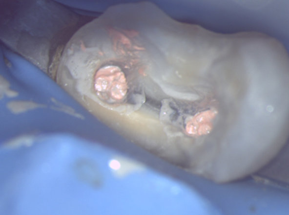
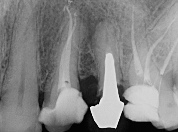
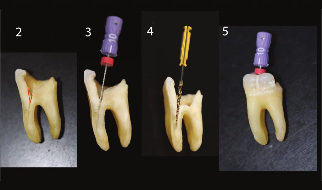
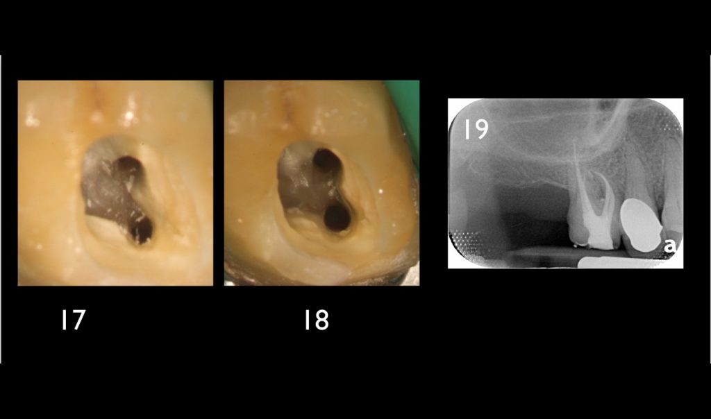
Dr Alberto DAGNA, Italy - 1/14 First upper left premolar.
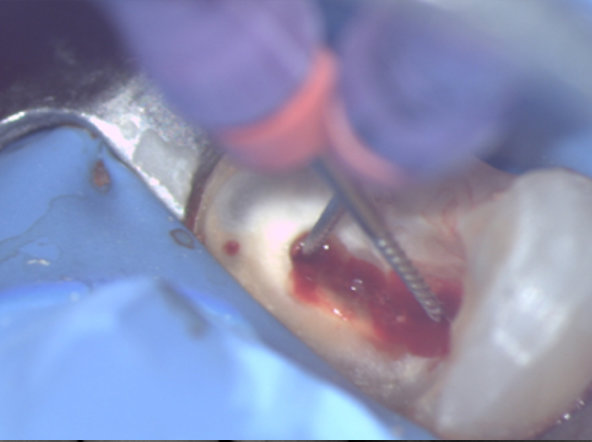
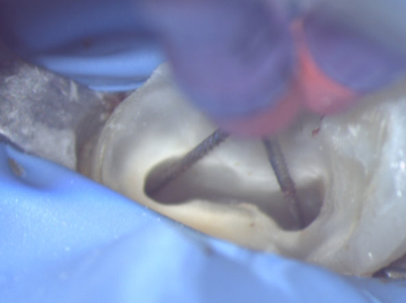
Dr Alberto DAGNA, Italy - 2/14 Access cavity and preliminary canal scouting with MMC files no.10.
Dr Alberto DAGNA, Italy - 3/14 Access cavity and preliminary canal scouting with MMC files no.10.
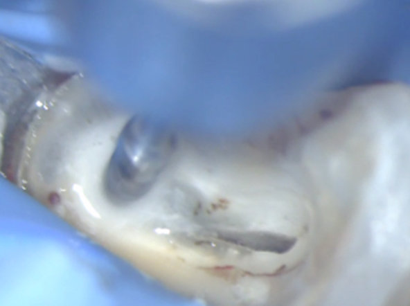
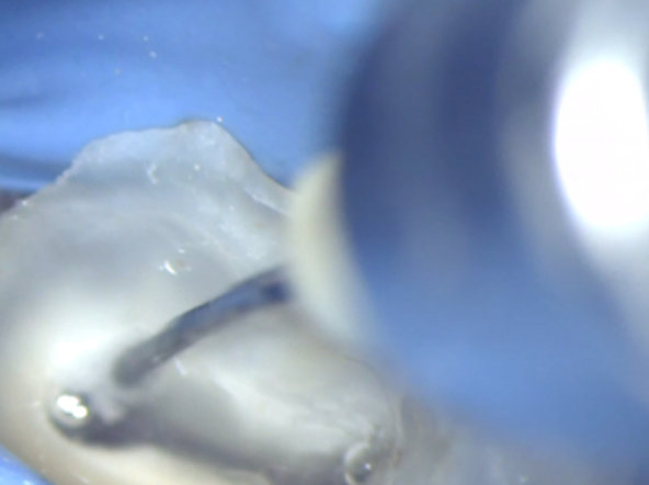
Dr Alberto DAGNA, Italy - 4/14 Orifices opening with ENDOFLARE.
Dr Alberto DAGNA, Italy - 5/14 Orifices opening with ENDOFLARE.
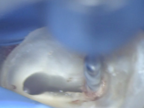
Dr Alberto DAGNA, Italy - 6/14 Orifices after the use of ENDOFLARE.
Dr Alberto DAGNA, Italy - 7/14 Orifices after the use of ENDOFLARE.
Dr Alberto DAGNA, Italy - 8/14 Preflaring using One G.
Dr Alberto DAGNA, Italy - 9/14 Preflaring using One G.
Dr Alberto DAGNA, Italy - 10/14 Scouting after preflaring Root canal shaping with One Shape
Dr Alberto DAGNA, Italy - 11/14 Scouting after preflaring Root canal shaping with One Shape
Dr Alberto DAGNA, Italy - 12/14 Final irrigation and obturation.
Dr Alberto DAGNA, Italy - 13/14 Final irrigation and obturation.
Dr Alberto DAGNA, Italy - 14/14 Post-operative x-ray.
One Flare
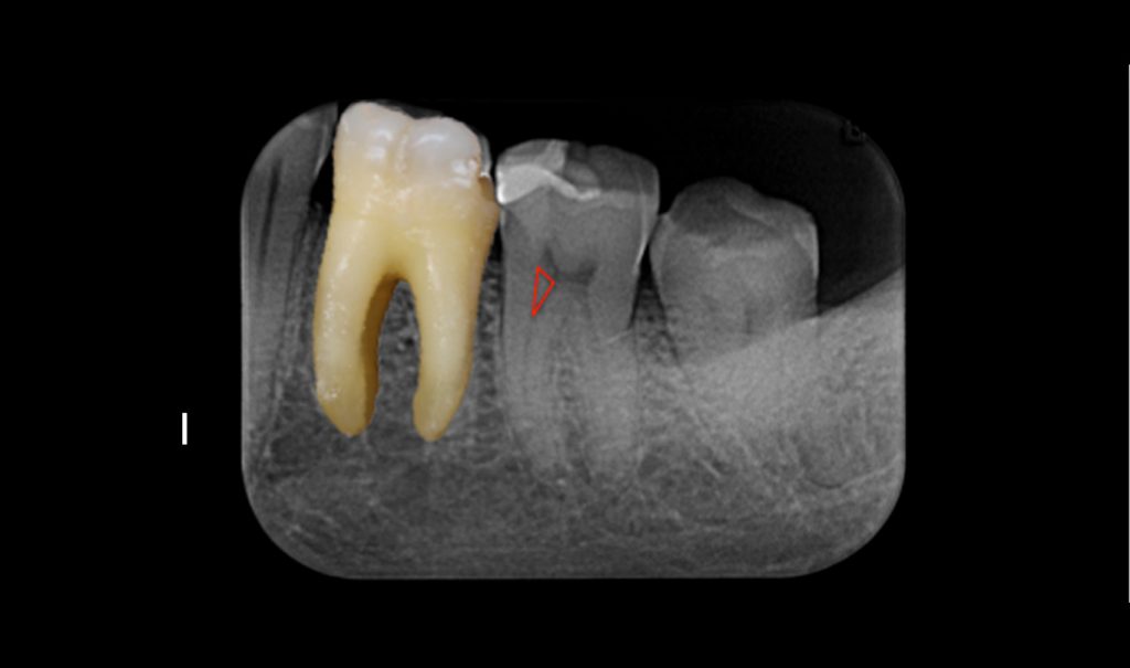
Schilder triangle: mineralization of the root canal entrance.
One Flare eliminates the Schilder triangle and the first millimeters of the pulp parenchyma to allow for a root canal shaping without excessive strain on the instruments after the determination of the apical preparation limit.
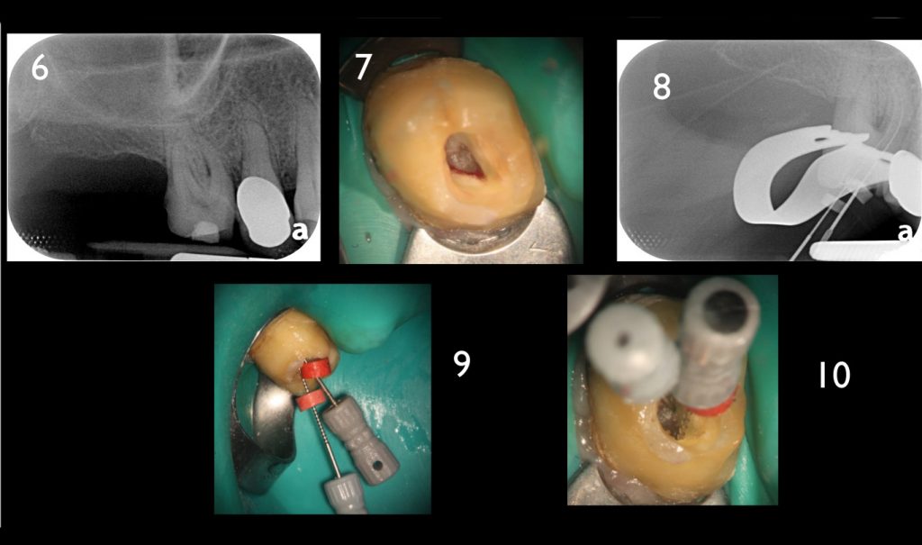
Initial x-ray, opening of the access cavity and x-ray with scouting file in the MB1 and MB2. The mineralization of the root canal entrance constrains the initial scouting.
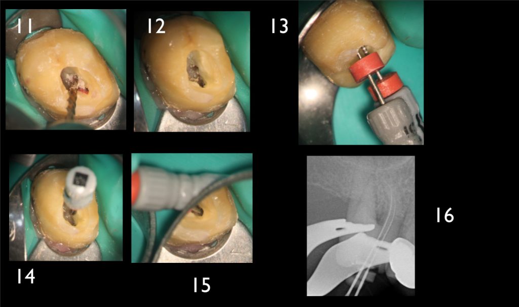
The flaring instrument One Flare eliminates the first millimeters of the pulp parenchyma, thus reducing the stress on the files which are realigned in the root canal, in order to facilitate the access to and the shaping at the apical preparation limit.
The reasoned preparation of the root canal entrance with the flaring instrument One Flare allows for safe endodontic shaping and cleaning while preserving the root canal’s homothety.
One Flare: SEM view of the profile and the tip, Prof. F. Diemer.
One Curve
One Curve & MM-Tooth
A video by Dr Filippo CARDINALI
One Curve in 1 min video
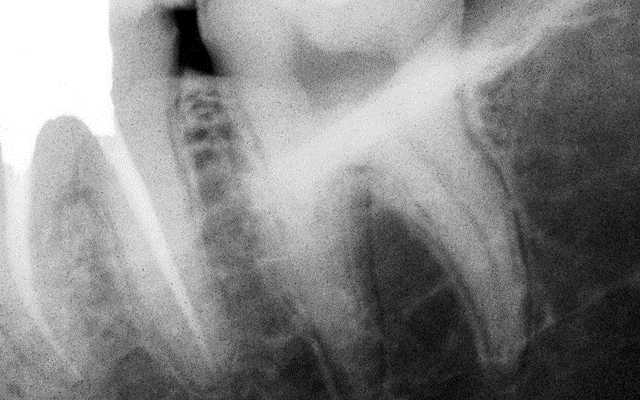
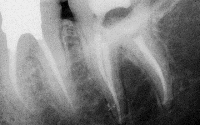
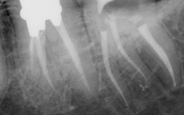
Dr Tara Mc MAHON, Belgium - 1/3 Preoperative x-ray Apical periodontitis, tooth n°37
Dr Tara Mc MAHON, Belgium - 2/3 Peroperative x-ray Removal of the old restauration and the cavity. Working length (WL) determination.
Dr Tara Mc MAHON, Belgium - 3/3 Post-operative x-ray Mesial canals: One Curve is used up to the first curvature, rinse. Second wave with One Curve down to the apex. Distal canal: One Curve direct to the apex, NaOCl 5,25% then EDTA 17%, final rinse with NaOCl before obturation.
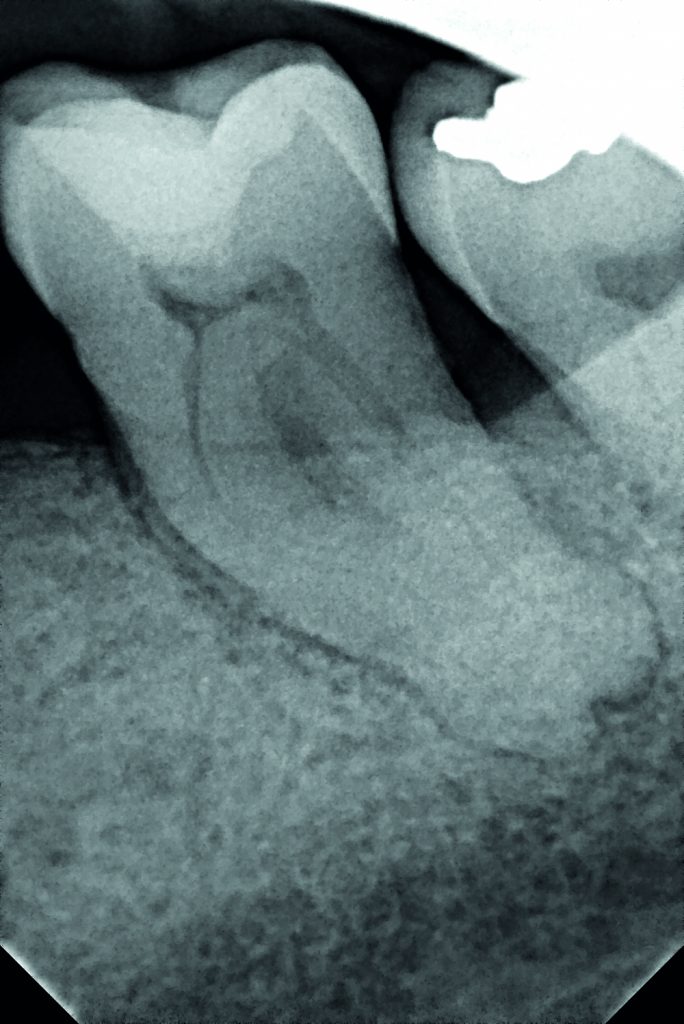
Pr K. BALTO, Saudi Arabia 1/3 Preoperative X-ray: tooth n°18 with irreversible pulpits and symptomatic apical periodontitis.
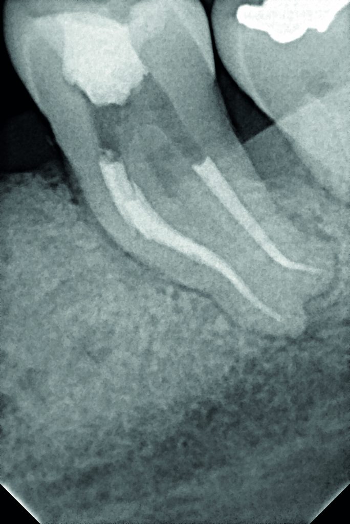
Pr K. BALTO, Saudi Arabia 2/3 Postoperative x-ray Glide path with One G, Preflaring with One flare, Shaping with One curve in three waves up to the apex.
Pr K. BALTO, Saudi Arabia 3/3 Postoperative x-ray Glide path with One G, Preflaring with One flare, Shaping with One curve in three waves up to the apex.
2Shape
2Shape in 2 min video
Clinical cases in video
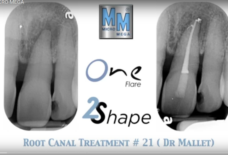
Dr Mallet #21
Clinical cases in video
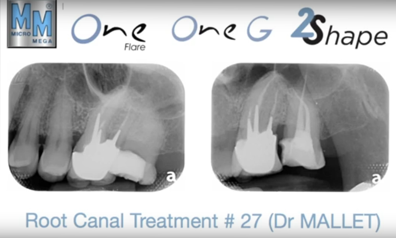
Dr Mallet #27
Clinical cases in video
Dr Cardinali
Pr. Franck Diemer, France - 1/2Preoperative x-ray of a patient’s necrotic 47 which received a crown reconstruction 6 months ago (glass ionomer cement + composite).
Pr. Franck Diemer, France - 2/2 Postoperative x-ray after shaping with 2Shape, apical finishing using F40 and obturation with pure thermomechanical condensation technique thanks to Revo Condensor.
Pr. Walid Nehme, Liban - 1/2 Preoperative x-ray of a 16 and a 17, both with deep caries and acute pulpitis.
Pr. Walid Nehme, Liban -2/2 Postoperative x-ray. Both molars were shaped using the 2Shape sequence. The apical finishing of the palatine canals was carried out with a F35 file.
Dr Jean-Philippe Mallet, France - 1/2 Preoperative x-ray: Upon diagnosis of pulp necrosis an initial endodontic treatment is indicated. The mesial root canals present a mineralized root canal lumen and a strong curvature and the distal canal an extremely curved apical third.
Dr Jean-Philippe Mallet, France - 2/2 Postoperative x-rays: The endodontic treatment is performed after scouting of the root canal with a diameter 10 hand file and securing of the mesial root canals using the NiTi file One G. Shaping was carried out with TS1 and TS2 to the working length, the MB2 canal was prepared using…
EndoUltra
EndoUltra in video
Dr. Riccardo Tonini, Italy - 1/2 Preoperative x-ray of tooth 35 affected by pulp necrosis.
Dr. Riccardo Tonini, Italy - 2/2 Postoperative x-ray after root canal shaping with the systems One G and 2Shape. 5 successive irrigation cycles with NaOCl activated with EndoUltra during 30 seconds. Final rinse with EDTA, activation and distilled water before obturation.
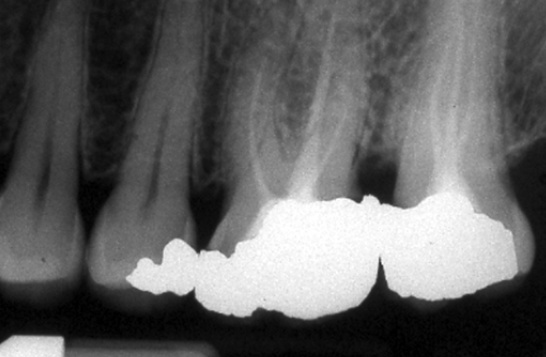
Dr. Rémy Balthazard, France - 1/2 Preoperative x-ray showing the necessity of an endodontic retreatment of the teeth no. 15, 16 and 17 as part of a global restoration.
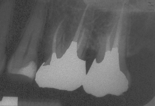
Dr. Rémy Balthazard, France - 2/2 Postoperative x-ray after root canal shaping with the systems One G and 2Shape. Irrigation with NaOCl at 3% and EDTA in a solution of 17%. Activation of the irrigation solutions with EndoUltra during 1 minute.
Dr. Riccardo Tonini, Italie - 1/2 X-ray of a tooth no. 34 at the end of retreatment. Presence of gutta percha residues in the retreated root canal.
Dr. Riccardo Tonini, Italie - 2/2 X-ray after activation of the NaOCl with EndoUltra in order to eliminate the gutta percha residues: 3 activation cycles of 30 seconds each, final rinse with distilled water before a new obturation.
Dr. Riccardo Tonini, Italy - 1/2 Preoperative x-ray of a tooth 37 affected by pulp necrosis.
Dr. Riccardo Tonini, Italy - 2/2 Postoperative x-ray after root canal shaping with 2Shape (TS1 and TS2), irrigation with NaOCl at 6% and EDTA 17%. Final irrigation protocol: 5 cycles with NaOCI which has been activated during 30 seconds using EndoUltraTM and one last activation cycle with EDTA. Rinse with distilled water.
R-Endo
1/2 ERT on 44. Preoperative X-ray.
2/2 Initial endodontic treatment on 45. Reproducibility ERT / Initial endodontic treatment is shown on these 2 premolars.
1/3 ERT on 26 and 27 Preoperative X-ray.
2/3 ERT on 26. Postoperative X-ray.
3/3 ERT on 26 and 27. Postoperative X-ray after 1 month.
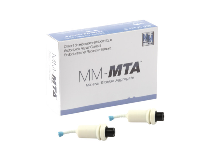
MM-MTA
MM-MTA in video
Dr Tara Mc MAHON, Belgique - 3/3 Radiographie post-opératoire Canaux mésiaux : One Curve jusqu’à la première courbure, rinçage. 2ème vague avec One Curve jusqu’à LT. Canal distal : One Curve directement à la LT, NaOCl 5,25% puis EDTA 17%, dernier rinçage NaOCl avant obturation.
Dr Tara Mc MAHON, Belgium - 3/3 Post-operative x-ray Mesial canals: One Curve is used up to the first curvature, rinse. Second wave with One Curve down to the apex. Distal canal: One Curve direct to the apex, NaOCl 5,25% then EDTA 17%, final rinse with NaOCl before obturation.
Dr Tara Mc MAHON, Belgien - 3/3 Postoperative Röntgenaufnahme Mesiale Kanäle: Verwendung von One Curve bis zur ersten Krümmung, Spülen. Zweite Verwendung von One Curve bis zur AL. Distaler Kanal: One Curve direkt bis zur AL, NaOCl 5,25 %, dann EDTA 17 %, letzte Spülung mit NaOCl vor der Obturation.
Pr K. BALTO, Arabie Saoudite 1/3 Radiographie pré-opératoire d'une dent n°18 présentant une pulpite irréversible et parodontite apicale symptomatique
Pr K. BALTO, Saudi Arabia 1/3 Preoperative X-ray: tooth n°18 with irreversible pulpits and symptomatic apical periodontitis.
Pr K. BALTO, Saudi Arabien 1/3 Präoperative Röntgenaufnahme eines Zahns Nr. 18 mit irreversibler Pulpitis und symptomatischer apikaler Parodontitis
Pr K. BALTO, Arabie Saoudite 2/3 Radiographie post-opératoire Exploration canalaire initiale, cathétérisme avec One G, évasement coronaire avec One Flare Mise en forme One Curve en trois vagues jusqu’à l’apex
Pr K. BALTO, Saudi Arabia 2/3 Postoperative x-ray Glide path with One G, Preflaring with One flare, Shaping with One curve in three waves up to the apex.
Pr K. BALTO, Saudi-Arabien 2/3 Postoperative Röntgenaufnahme Sondierung des Wurzelkanals, Gleitpfad mit One G, koronale Erweiterung mit One Flare, Wurzelkanalaufbereitung mit One Curve in drei Etappen bis zum Apex.
Pr K. BALTO, Arabie Saoudite 3/3 Radiographie post-opératoire Exploration canalaire initiale, cathétérisme avec One G, évasement coronaire avec One Flare Mise en forme One Curve en trois vagues jusqu’à l’apex
Pr K. BALTO, Saudi Arabia 3/3 Postoperative x-ray Glide path with One G, Preflaring with One flare, Shaping with One curve in three waves up to the apex.
Pr K. BALTO, Saudi-Arabien 3/3 Postoperative Röntgenaufnahme Sondierung des Wurzelkanals, Gleitpfad mit One G, koronale Erweiterung mit One Flare, Wurzelkanalaufbereitung mit One Curve in drei Etappen bis zum Apex.
Fusion de Micro-Mega, SCICAN et du groupe COLTENE
October 2018: merge of Micro-Mega, SCICAN and COLTENE